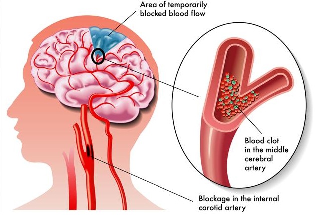
A stroke occurs when blood flow is cut off from part of the brain. In the same way that a person suffering a loss of blood to the heart can be said to be having a “heart attack,” a person with a loss of blood to the brain can be said to be having a “brain attack.” There are two kinds of stroke, hemorrhagic and ischemic. Hemorrhagic strokes are caused by bleeding within the brain. Ischemic strokes, which are far more common, are caused by a blockage of blood flow in an artery in the head or neck leading to the brain. Some ischemic strokes are due to stenosis, or narrowing of arteries due to the build up of plaque, fatty deposits and blood clots along the artery wall. A vascular disease that can cause stenosis is atherosclerosis, in which deposits of plaque build-up along the inner wall of large and medium-sized arteries, decreasing blood flow. Atherosclerosis in the carotid arteries, two large arteries in the neck that carry blood to the brain, is a major risk factor for ischemic stroke.
Our Service
CAROTID ARTERY DISEASES
Blockage in the carotid arteries in the neck is a common and important cause for the development of brain stroke or paralysis, a potentially disabling and often life-threatening condition. The role of a vascular specialist in removing this blockage is vital for the prevention of stroke. The causes or risk factors for blockage in the carotid artery are similar to those for blockages in the heart or leg artery.
What is a stroke?

What are the symptoms of a stroke?
Weakness or num bness on one side (leg, arm or face], slurred speech or visual disturbances are the usual presenting symptoms of a mini -stroke. Even if these events last for a few minutes or hours, they could be warning signs of carotid blockage and an impending stroke.
These symptoms need to be evaluated by a carotid Doppler test, which is a simple non – i nvasive sonography of the neck. A confirmatory angiography or CT angiography is performed if an intervention is planned.
Additionally, patients with high blood pressure, diabetes, cholesterol or past heart problems should undergo a baseline carotid Doppler test

WHAT ARE THE TREATMENT OPTIONS?
Minor blockages (less than 50%] are treated with blood thinners and cholesterol-controlling medicines. If the blockage is more than 50%, surgery or angioplasty with stenting is recommended.
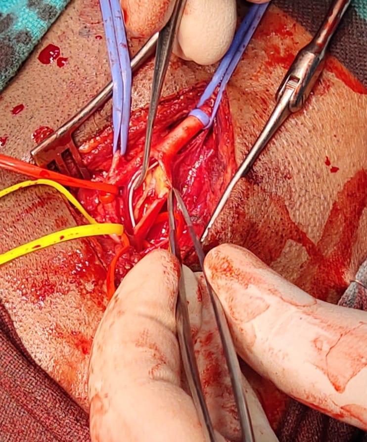
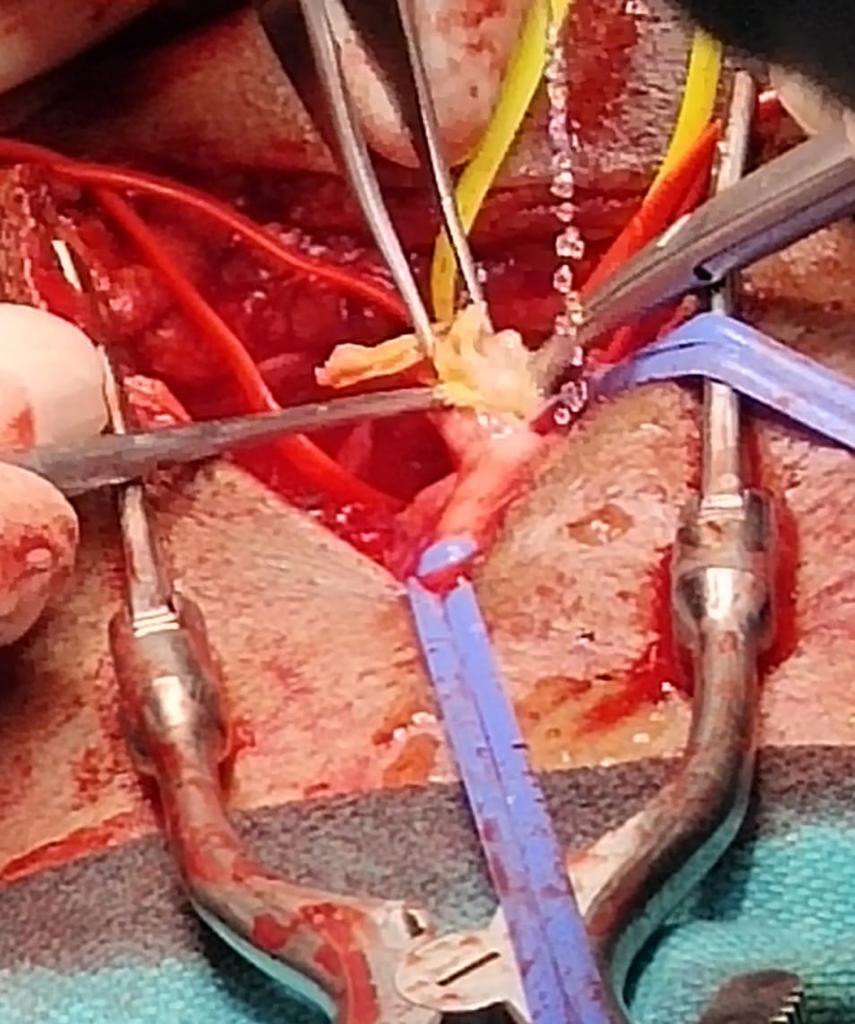
Carotid endarterectomy is a special surgical procedure for the removal of the carotid artery blockage. We routinely perform this procedure under a loco – regional anesthesia for the ease in intra-operative neurological monitoring. The
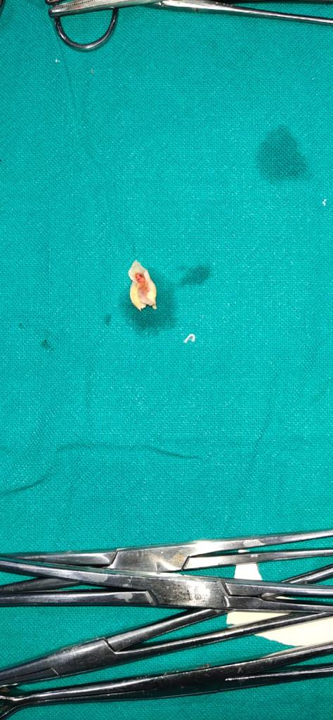
artery is opened and the plaque carefully removed. During the surgery, we use a shunt to maintain blood flow to the brain.
The artery is then repaired using a vein or graft patch to prevent narrowing.


What is “best medical therapy” for stroke prevention?
The mainstay of stroke prevention is risk factor management
How is carotid artery disease diagnosed?
In some cases, the disease can be detected during a normal checkup by a physician. In other cases further testing is needed. Some of the tests a physician can use or order include ultrasound imaging, arteriography, and magnetic resonance angiography (MRA). Frequently these procedures are carried out in a stepwise fashion: from a doctor’s evaluation of signs and symptoms to ultrasound, MRA, and arteriography for increasingly difficult cases.
History and physical exam
A doctor will ask about symptoms of a stroke such as numbness or muscle weakness, speech or vision difficulties, or lightheadedness. Using a stethoscope, a doctor may hear a rushing sound, called a bruit (pronounced “broo-ee”), in the carotid artery. Unfortunately, dangerous levels of disease sometimes fail to make a sound, and some blockages with a low risk can make the same sound.
Ultrasound imaging
This is a painless, noninvasive test in which sound waves above the range of human hearing are sent into the neck. Echoes bounce off the moving blood and the tissue in the artery and can be formed into an image. Ultrasound is fast, risk-free, relatively inexpensive, and painless compared to MRA and arteriography.
Arteriography
This can be used to confirm the findings of ultrasound imaging which can be uncertain in some cases. Arteriography is an X-ray of the carotid artery taken when a special dye is injected into the artery. A burning sensation may be felt when the dye is injected. An arteriogram is more expensive and carries its own small risk of causing a stroke.
Magnetic Resonance Angiography (MRA)
This is a new imaging technique that avoids most of the risks associated with arteriography. An MRA is a type of image that uses magnetism instead of X-rays to create an image of the carotid arteries.