Congenital vascular malformations (often termed as birthmarks) are one of the most challenging subgroups of diseases treated by vascular surgeons and intervention lists. There are a number of controversies and differences in the diagnosis or treatment of vascular malformation.
CLASSIFICATION
Currently, International ISSVA classification system is widely accepted and utilized to categorize vascular anomalies into two basic types:
Vasoproliferative or vascular neoplasm’s such as hemangioma
Developmental vascular abnormalities called congenital vascular malformations (CVMs)
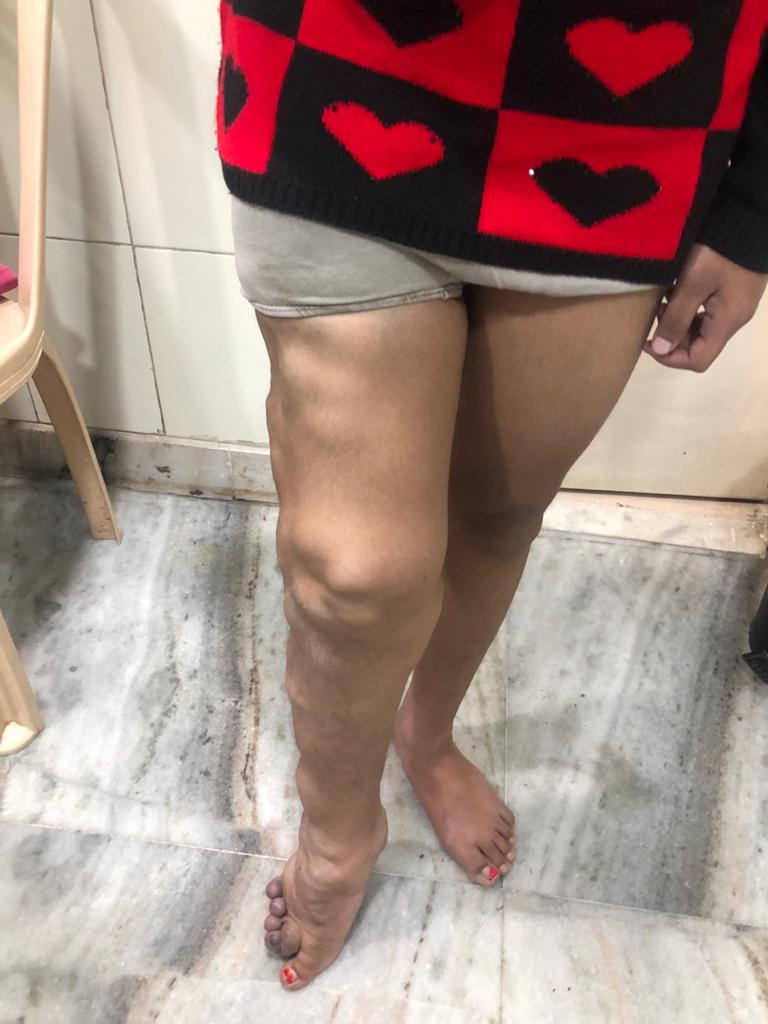
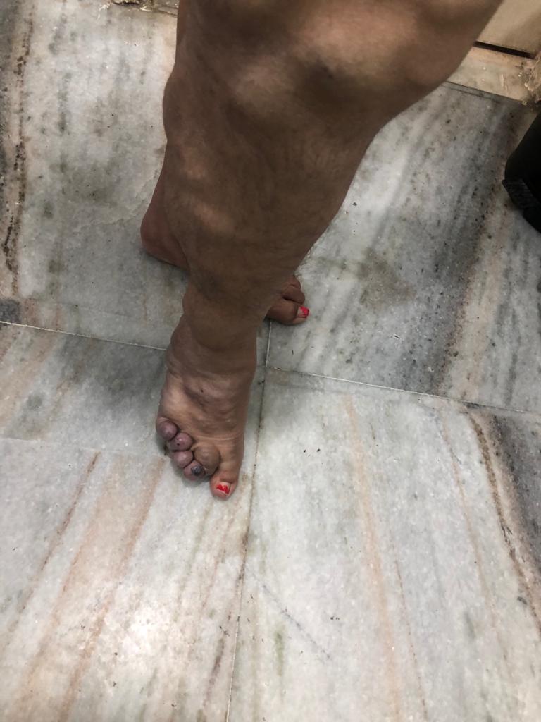
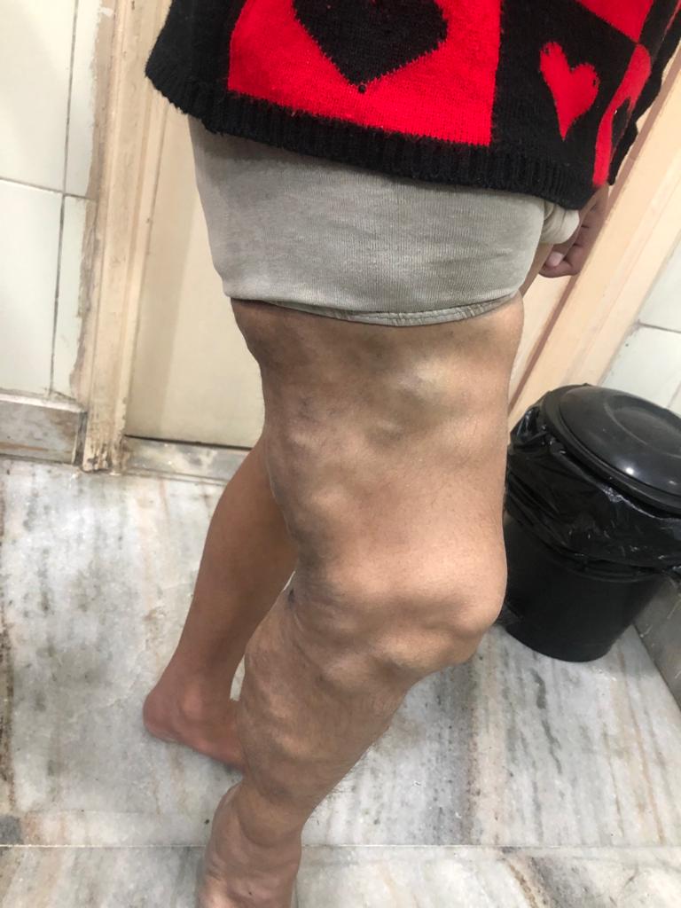
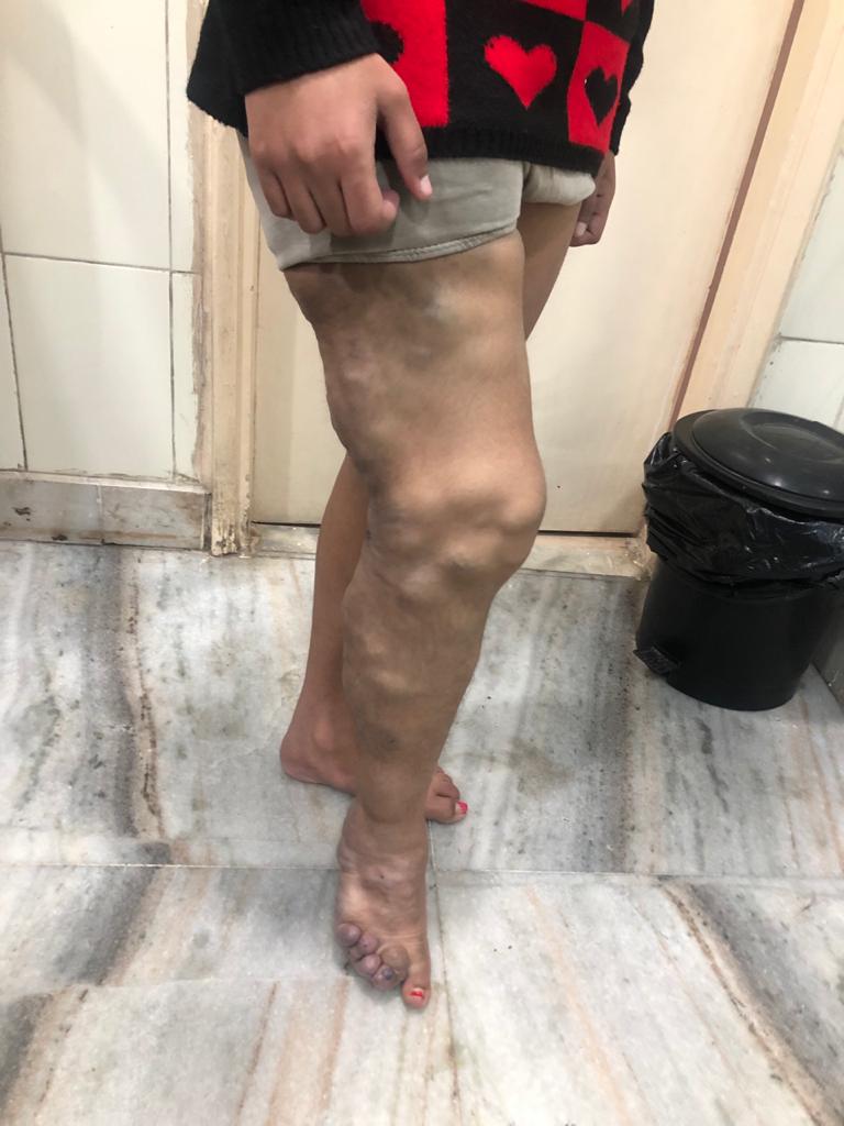
CVMs are a large group which include venous (VM), arterial (AM), capillary (CM), and lymphatic (LM), as well as arteriovenous (AVM) malformations.
The clinical importance of classification is important to reduce the confusion in treatment principles.
CLINICAL FEATURES
Infantile hemangiomas which are commonly seen after birth and appear as strawberry mark do not require immediate intervention and can undergo spontaneous involution (shrinkage) or recovery (60%–70% involution by the age of two years and 90% by the age of 7 years).
Venous malformations may appear as bluish, soft compressible masses (subcutaneous or mucosal) without any bruit (spread)/thrill. The presence of thrill is typical of AVM. Lymphatic malformations may present as cystic variants (lymphangioma) or soft but non-compressible masses (microcystic variety).
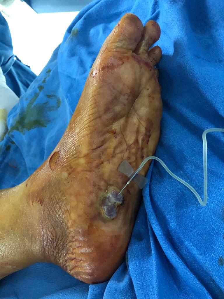
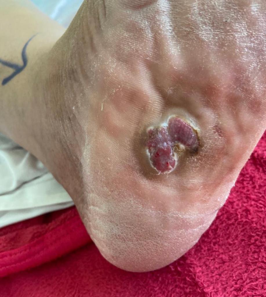
Some vascular malformations present with swelling, pain, ulcer or bleeding depending on the location and extent of the malformation.
DIAGNOSIS
After a clinical examination, Doppler ultrasound is the next diagnostic step, following which an MRI or a CT angiography may be performed as per your doctors’ advice.
GENERAL TREATMENT PRINCIPLES
Not all CVMs need aggressive They can be managed conservatively using compression stockings, if possible.
They are slowly progressive and may show sudden increase during puberty or post
CVMs are best managed by a multidisciplinary approach utilising a combination of nonsurgical, surgical and endovascular methods
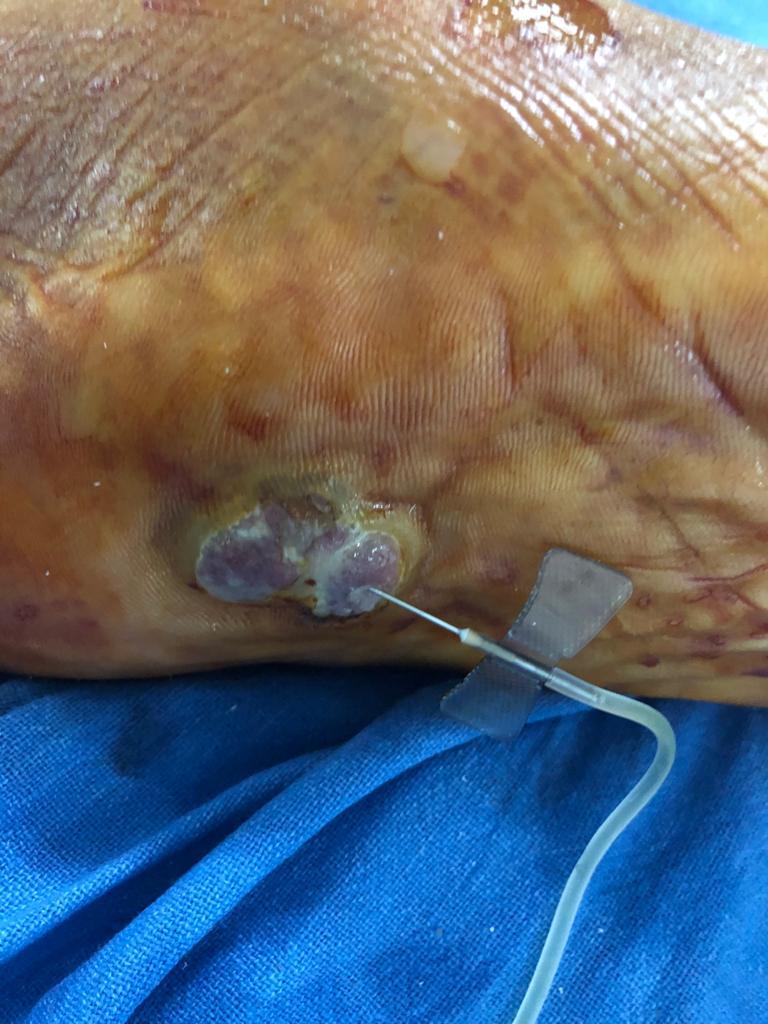
SCLEROTHERAPY
This is the mainstay of treatment of most CVMs, in which single or multiple sessions of injections of sclerosant solution (STS, bleomycin or alcohol) are given in the lesion under Doppler or fluoroscopy guidance.
Although initially patient can feel hardness or bruising at the injection site, this treatment can eventually help to reduce the size of the swelling.
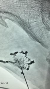
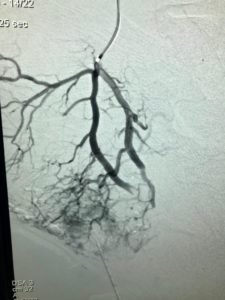
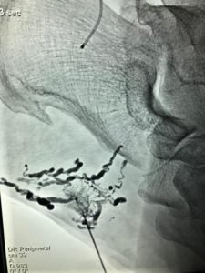
ANGIOEMBOLISATION
This minimally invasive complex treatment method is mainly used for AVM. In this technique, fine catheters are introduced within the blood vessels and advanced to the centre of the AVM (nidus) with placement of liquid embolic agents (alcohol, glue, Onyx) to help in the reduction of the blood flow.
Multiple sessions of angioembolisation are often required to achieve significant reduction in the size of the AVM.
SURGERY
Surgery is usually restricted to treat lesions where total excision would be possible without causing local damage to important neurologist or vascular tissues.
Some patients with AVM may require angio embolization followed by surgical excision to provide complete cure.