Table of Contents
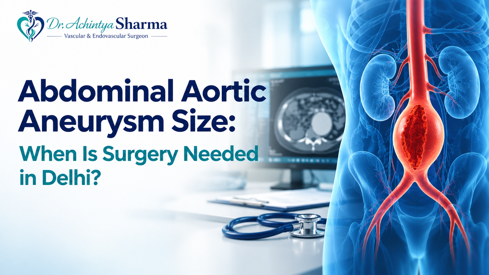
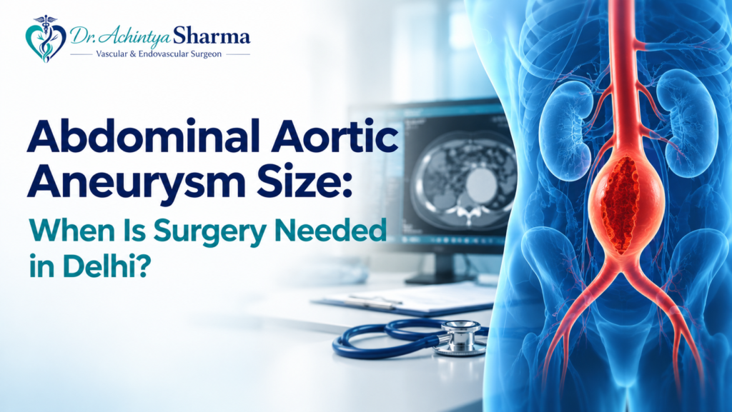
An abdominal aortic aneurysm (AAA) occurs when a portion of the aorta inside the abdomen becomes abnormally wide. This blood vessel supplies blood to the lower part of the body. As the affected area enlarges, the vessel wall may become weaker, creating a risk of rupture in some patients. Many people do not experience noticeable symptoms, even when an aneurysm is present.
The abdominal aortic aneurysm size is an important measurement used to evaluate the condition and plan appropriate care. Doctors also consider how quickly it is growing, whether it causes symptoms and whether the patient can safely undergo a procedure.
This article explains aneurysm measurements, available investigations and treatment approaches for people seeking vascular care in Delhi.
The information below is for educational purposes and should not replace advice from a qualified medical professional.
Understanding Abdominal Aortic Aneurysm Measurements
An aneurysm measurement represents the widest diameter of the affected section of the aorta. It is generally recorded in centimetres and assessed through medical imaging.
A diameter of 3.0 cm or greater is commonly used to define an abdominal aortic aneurysm. However, the measurement must be interpreted alongside the patient’s medical history and other clinical findings.
Ultrasound is frequently used to identify the condition and track changes over time. When a more detailed view of the blood vessels is required, a doctor may recommend CT angiography. This investigation can be particularly useful when evaluating the suitability of a surgical procedure.
Since an aneurysm may enlarge without causing pain, following the recommended monitoring schedule is essential.
Abdominal Aortic Aneurysm Size Chart
The table below provides a general reference for understanding different measurement ranges.
| Diameter | Common management approach |
| Below 3.0 cm | Usually falls outside the definition of an aneurysm |
| 3.0–3.9 cm | Usually monitored periodically |
| 4.0–4.9 cm | Continued surveillance is important |
| 5.0–5.4 cm | Specialist evaluation helps determine the next steps |
| 5.5 cm or more | Repair may be recommended for suitable patients |
These ranges are intended as general guidance. They do not determine the treatment required by an individual. The threshold for considering repair may differ according to sex, symptoms, aneurysm growth and the clinical guideline used.
A specialist should review the complete scan report before recommending further management.
When Should an Abdominal Aortic Aneurysm Be Repaired?
The decision to operate depends on whether the expected benefit of repair outweighs the risks of the procedure. Some patients need regular scans, while others may require earlier intervention.
NICE guidance advises considering repair when an unruptured aneurysm produces symptoms, reaches 5.5 cm in diameter or measures more than 4.0 cm and increases by over 1 cm within 12 months.
Treatment recommendations can vary across guidelines and individual clinical circumstances. The decision should therefore be made after a specialist assessment.
Important considerations include:
- Size: Larger aneurysms generally have a greater risk of rupture.
- Expansion: Faster enlargement may indicate the need for an earlier review.
- Symptoms: Pain associated with the aneurysm may affect the urgency of treatment.
- Medical fitness: Other health conditions can influence the risks of an operation.
- Vascular anatomy: The position and shape of the affected artery help determine the appropriate procedure.
For many patients with smaller, symptom-free aneurysms, regular imaging remains a suitable approach.
Tests Used for Diagnosis and Follow-Up
Medical investigations help establish the diagnosis and identify changes that may affect treatment decisions.
Ultrasound Examination
Ultrasound uses sound waves to create images of the abdominal aorta. It is commonly used for initial assessment and repeated measurements because it is non-invasive and does not use ionising radiation.
CT Angiography
This scan provides detailed images of the aorta and nearby blood vessels. It can help doctors assess the structure of the aneurysm and plan an appropriate repair technique when intervention is being considered.
Regular Medical Reviews
The recommended interval between scans depends on the aneurysm’s diameter, growth and other risk factors. Keeping earlier reports available helps the treating doctor compare measurements and identify important changes.
Patients should continue scheduled follow-up even when they have no symptoms.
EVAR and Open Surgery: What Is the Difference?
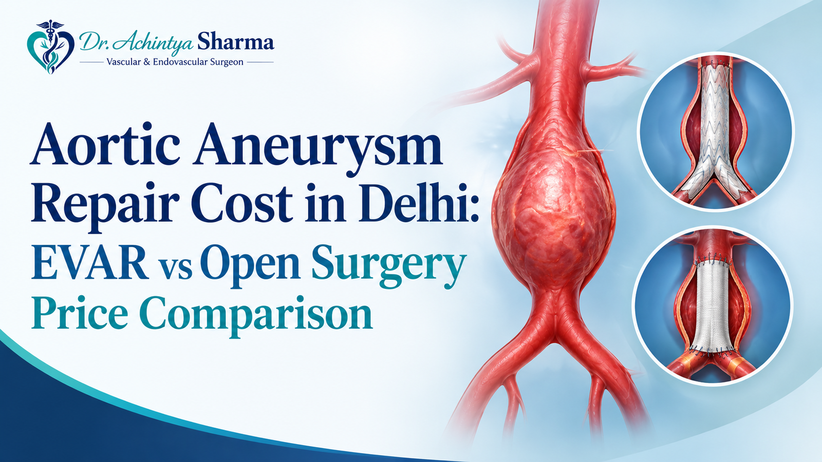
When an aneurysm requires repair, doctors generally consider two main approaches: endovascular aneurysm repair (EVAR) and open surgical repair.
Endovascular Aneurysm Repair
EVAR involves placing a specialised stent graft inside the aorta through blood vessels, usually accessed through the groin. The graft creates a supported channel for blood flow and helps reduce pressure on the weakened section.
Because the procedure is less invasive, the initial recovery is often shorter than with open surgery. However, not every aneurysm is suitable for EVAR. Patients also require follow-up imaging, and some may need additional treatment later.
Open Surgical Repair
Open repair involves an abdominal incision to reach the affected artery. The surgeon uses a graft to replace or reinforce the weakened section of the aorta.
Recovery generally takes longer than after EVAR. However, open surgery may be appropriate when the aneurysm’s structure does not allow a suitable endovascular repair.
| Consideration | EVAR | Open repair |
| Procedure | Stent graft placed through blood vessels | Surgery performed through an abdominal incision |
| Initial recovery | Often shorter | Usually longer |
| Eligibility | Depends on aneurysm anatomy | Considered according to anatomy and patient factors |
| Aftercare | Continued imaging is required | Regular clinical follow-up is important |
The choice between these procedures depends on anatomy, overall health, expected outcomes and patient preferences. A vascular surgeon can explain the risks and benefits of each option.
Risk Factors for Developing an Aneurysm
Several factors are associated with an increased likelihood of developing this condition. These include:
- Older age.
- Smoking or a history of smoking.
- Family history of abdominal aortic aneurysm.
- High blood pressure and other cardiovascular risk factors.
- Certain inherited disorders that affect the blood vessels.
People who may be at increased risk can ask their healthcare professional whether screening is appropriate. Recommendations depend on personal risk factors and medical history.
Finding Abdominal Aortic Aneurysm Treatment in Delhi
People seeking an abdominal aortic aneurysm specialist in Delhi can arrange an assessment with a qualified vascular and endovascular surgeon. A consultation can help clarify the scan findings, estimate potential risks and establish whether observation or repair should be considered.
Visit Dr. Achintya Sharma’s website for information about aortic aneurysm assessment and treatment options.
For a productive appointment, carry previous imaging reports, current medication details and relevant medical records. You may also want to ask the specialist:
- What do my latest scan measurements indicate?
- Has the aneurysm changed since my previous examination?
- When should I undergo another scan?
- What factors would make surgery necessary?
- Which repair method may be suitable for me?
- What complications and follow-up requirements should I understand?
An appropriate care plan should be based on individual findings rather than a single measurement.
Warning Signs That Need Immediate Attention
A rupture can cause severe internal bleeding and requires emergency treatment. Sudden, intense pain in the abdomen or back, particularly when accompanied by fainting, collapse, dizziness or marked weakness, should never be ignored.
Seek emergency medical care immediately if these symptoms occur. Do not wait for a routine appointment or a scheduled scan.
Frequently Asked Questions
1. What diameter is considered normal for the abdominal aorta?
A diameter below 3.0 cm is generally not classified as an abdominal aortic aneurysm. A measurement of 3.0 cm or more commonly meets the definition, subject to clinical interpretation.
2. Does a 4 cm aneurysm always require surgery?
No. A stable aneurysm of this size without symptoms is often managed through regular imaging. The monitoring plan depends on individual clinical findings.
3. Is surgery necessary when the measurement reaches 5 cm?
Not in every case. Doctors consider symptoms, growth, sex, anatomy and overall health before deciding whether repair or continued monitoring is appropriate.
4. Can an aneurysm remain unnoticed?
Yes. Many aneurysms do not produce obvious symptoms. Recommended scans are important because changes may occur without the patient noticing them.
5. Which investigation is used to examine an aneurysm?
Ultrasound is commonly used for diagnosis and monitoring. CT angiography may provide additional anatomical details when further evaluation or treatment planning is needed.
6. Can lifestyle changes reverse an aneurysm?
Lifestyle measures cannot remove an existing aneurysm. Stopping smoking and controlling blood pressure can support cardiovascular health, but they do not replace prescribed monitoring or treatment.
Conclusion
An abdominal aortic aneurysm requires careful monitoring and appropriate medical attention. If you have been diagnosed with an aneurysm or are concerned about its size, growth or potential complications, consult a qualified vascular specialist to understand your condition and available treatment options. Early evaluation can help determine whether regular monitoring or surgical repair may be appropriate.
Don’t wait for symptoms to worsen. Contact a vascular specialist in Delhi today to discuss your scan reports and explore suitable treatment options for your condition.